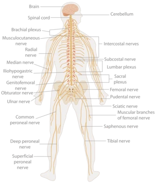
Human body
The entire structure of a human being.
The human body is a complete structure made of trillions of cells, which combine to form tissues, then organs, and finally organ systems. Its external parts include the head, hair, neck, torso (made up of the thorax and abdomen), genitals, arms, hands, legs, and feet. Inside, it contains organs, teeth, bones, muscles, tendons, ligaments, blood vessels and blood, lymphatic vessels and lymph. The body is studied through anatomy, physiology, histology, and embryology, and it varies anatomically in known ways. Physiology looks at how systems and organs function, with many systems working together to maintain homeostasis—keeping safe levels of substances like sugar, iron, and oxygen in the blood. Health professionals, physiologists, anatomists, and artists study the body to aid their work.
The body is built from elements such as hydrogen, oxygen, carbon, calcium, and phosphorus, found in trillions of cells and non-cellular parts. An adult male body is about 60% water, totaling roughly 42 liters. This includes around 19 liters of extracellular fluid (about 3.2 liters of blood plasma and 8.4 liters of interstitial fluid) and about 23 liters inside cells. The water’s content, acidity, and composition are carefully regulated. Outside cells, the main electrolytes are sodium and chloride; inside cells, they are potassium and other phosphates.
There are trillions of cells—the basic unit of life. At maturity, the body has about 30 trillion human cells and 38 trillion bacteria, a count based on summing all organ and cell types. The skin hosts billions of commensal organisms and immune cells. Not all body parts are made of cells; cells sit in an extracellular matrix of proteins like collagen, surrounded by fluids. Each cell experiences tens of thousands of DNA damages daily, which can block genome replication or transcription. If unrepaired or repaired incorrectly, these damages may cause mutations or other genome changes that threaten cell survival.
Cells function because of DNA, which sits in the cell nucleus. Parts of DNA are copied and sent via RNA to the cell body, where RNA creates proteins. Proteins form the basis for cells, their activity, and products, dictating cell function and gene expression. Cells self-regulate by controlling protein amounts. However, not all cells have DNA—mature red blood cells lose their nucleus as they mature.
The body has many tissue types, defined as cells with specialized functions. The study of tissues is histology, often done with a microscope. There are four main tissue types: lining cells (epithelia), connective tissue, nerve tissue, and muscle tissue. Lining cells cover surfaces exposed to the outside world or the gastrointestinal tract (epithelia) or internal cavities (endothelium). They come in various shapes—single layers of flat cells, cells with tiny beating cilia in the lungs, or column-like cells lining the stomach. Endothelial cells line internal cavities like blood vessels and glands. These lining cells regulate what passes through, protect internal structures, and act as sensory surfaces.
Organs are structured collections of cells with specific functions, mostly inside the body except for the skin. Examples include the heart, lungs, and liver. Many organs sit in body cavities, such as the abdomen (housing the stomach) and the pleura (containing the lungs).
The heart is located in the thoracic cavity between the lungs, slightly to the left. It is surrounded by the pericardium, which holds it in place in the mediastinum, protects it from blunt trauma and infection, and lubricates its movement with pericardial fluid. The heart pumps blood around the body, transporting oxygen, nutrients, waste, hormones, and white blood cells. It has two atria and two ventricles. The atria allow uninterrupted venous blood flow during ventricular systole, letting enough blood enter the ventricles during atrial systole, boosting cardiac output by about 75%. The ventricles pump blood to the lungs (right ventricle) and the rest of the body (left ventricle). The heart has an electrical conduction system that controls muscle contraction and relaxation. It starts in the sinoatrial node, travels through the atria to pump blood into the ventricles, then goes to the atrioventricular node, where the signal slows slightly to let the ventricles fill before pumping out and restarting the cycle. Coronary artery disease is the leading cause of death worldwide, accounting for 16% of all deaths. It results from plaque buildup in the coronary arteries, which can narrow them so much that not enough blood reaches the myocardium—a condition called myocardial infarction or heart attack—potentially leading to heart failure or cardiac arrest.
- field
- Anatomy, physiology, histology, embryology
- known_for
- Complex organization from cells to organ systems; maintenance of homeostasis
Lore & Background
The human body consists of a head, hair, neck, torso (including thorax and abdomen), genitals, arms, hands, legs, and feet. Internally, it includes organs, teeth, bones, muscles, tendons, ligaments, blood vessels and blood, lymphatic vessels and lymph. The body is composed of elements such as hydrogen, oxygen, carbon, calcium, and phosphorus, residing in trillions of cells and non-cellular components. The adult male body is about 60% water, with carefully maintained content, acidity, and composition inside and outside cells.
Reader's Guide
The human body is the fundamental subject of anatomy, physiology, histology, and embryology. Its study enables health professionals to diagnose and treat conditions, physiologists to understand function, anatomists to map structure, and artists to depict the human form accurately. The body's systems, such as the circulatory and digestive systems, interact to maintain homeostasis, with safe levels of substances like sugar, iron, and oxygen in the blood. Understanding the body's composition, from cells to organ systems, is essential for medical science and biological research.
Did You Know?
- The adult male body is about 60% total body water content, some 42 litres.
- At maturity, there are roughly 30 trillion human cells and 38 trillion bacteria in the body.
- Coronary artery disease is the leading cause of death worldwide, making up 16% of all deaths.
- The heart is composed of two atria and two ventricles.
The Hierarchical Architecture of the Human Body
The human body is organized in a nested hierarchy that moves from the smallest building blocks to the largest functional units. At the foundation sit individual cells and connective tissue, which assemble into tissues. Those tissues then combine to form organs, and organs work together within biological systems. This layered organization means that understanding the body requires moving between scales—examining a single cell under a microscope, then stepping back to see how that cell's tissue contributes to an organ's function, and finally appreciating how that organ fits into a whole system. This is why anatomy is split into two major branches: gross anatomy, which deals with structures visible to the naked eye, and microscopic anatomy, which requires magnification to reveal histology (tissue organization) and cytology (cell structure). The complementary fields of physiology and biochemistry round out the picture, addressing function and chemistry respectively, and together they form the core basic medical sciences taught to first-year medical students.
Evolutionary Echoes in Human Structure
Human anatomy is not an isolated design; it carries deep evolutionary signatures that link us to all vertebrates. Much of the body retains an ancient segmental pattern—repeating basic units that are particularly visible in the vertebral column and the ribcage. This pattern can be traced back to very early embryonic development, revealing that the architecture we see in an adult was laid down in the earliest stages of growth. Because of these shared roots, human anatomy is closely intertwined with embryology, comparative anatomy, and comparative embryology. Studying how a human embryo develops, or how the same segmental logic appears in fish, reptiles, and birds, illuminates why our own structures are arranged the way they are. The history of anatomy itself reflects this deepening understanding: over centuries, the field has moved from simple observation toward a continually refined comprehension of how organs and structures relate to one another and to the broader evolutionary story.
Anatomy as a Foundation for Visual Arts
Long before medical imaging or digital rendering, artists turned to the human body's internal architecture to make their figures feel alive. Gross anatomy has become a cornerstone of visual arts because understanding how muscles and bones shift, deform, and interact during movement is essential to drawing, painting, or animating a convincing human figure. Leonardo da Vinci is a landmark example: he deliberately dissected and studied the human body to improve his artistic technique, and in doing so he simultaneously advanced the scientific understanding of anatomy and its representation on canvas. Today, dedicated guides such as Human Anatomy for Artists: The Elements of Form continue this tradition, offering painters and illustrators a structured way to render the body with anatomical accuracy. The complexity of a living organism means that artists, like anatomists, must think in layers—moving from the smallest structural details to the overall relationship between organs and the body's surface.
The Evolution of Anatomical Study and Teaching
The methods used to study the human body have transformed dramatically across the centuries. Early anatomists relied on examining animals, then progressed to dissecting fresh and preserved cadavers. The twentieth century brought technologically complex techniques that expanded what could be observed and understood. In modern medical education, students of medicine, dentistry, physiotherapy, nursing, paramedicine, and radiography learn gross and microscopic anatomy through a blend of models, skeletons, textbooks, diagrams, photographs, lectures, and tutorials. Medical and dental students additionally gain hands-on experience by dissecting and inspecting cadavers, while histology is reinforced by examining prepared slides under a microscope. Teaching can follow a regional approach—grouping structures by body area such as the head and neck, thorax, abdomen, pelvis, or limbs—or a systemic approach organized around functional systems like the nervous or respiratory. Gray's Anatomy, the field's landmark textbook, recently shifted from a systems format to a regional one, aligning with contemporary pedagogical preferences.
Frequently Asked Questions
What is the Human body in the Human Anatomy 25-38 series?
The Human body is the central subject of the entry, representing the full structural and functional makeup of a human being. It is built from countless specialized cells that assemble into tissues, organs, and interconnected organ systems.
What are the Human body's signature abilities?
Its two most celebrated traits are the layered complexity that runs from individual cells all the way up to whole organ systems, and its relentless drive to maintain internal stability (homeostasis) despite external changes.
Who are the main characters that interact with the Human body?
Health professionals, physiologists, anatomists, and artists are the recurring 'supporting cast' who study, treat, or depict the body to carry out their respective work.
Which fields does the Human body span across the series?
The entry draws on anatomy, physiology, histology, and embryology, giving the body a multi-disciplinary identity rather than belonging to just one specialty.
Why is the Human body considered the keystone of the 25-38 entry?
Because every other topic in the chapter ultimately traces back to how cells, tissues, and organs cooperate to keep a living person functional and balanced, making it the foundational reference point for the whole series.
More in Human Anatomy 1-24
Spotted an error? Know more?
This is a living reference — every entry is fact-audited, and reader corrections feed straight into our audit queue. Suggest an edit · See this site's audit record